
If you’ve been dealing with dry, irritated, or watery eyes, your optometrist could recommend something called a meibography. It may sound technical, but it’s simply an imaging technique used to look closely at a specific part of your eyelid: the meibomian glands.
These tiny glands don’t get much attention outside the exam room, but they play a huge role in your daily eye comfort. When they aren’t working well, your eyes can feel gritty, sore, or tired; and that’s where meibography comes in.
Why the Meibomian Glands Matter
Oil’s Role in Tear Stability
Along the edge of your upper and lower eyelids are rows of small oil glands. These are called meibomian glands, and their job is to release oil into your tears every time you blink. This oil helps keep your tears from evaporating too quickly.
When Things Go Wrong
When the glands become blocked or stop functioning properly, your tears can’t stay on the surface of your eye for long. That leads to dryness, irritation, and sometimes even blurry vision.
This condition is called meibomian gland dysfunction (MGD), and it’s one of the most common causes of dry eye disease. But because the problem lies inside your eyelid, it isn’t something that can be spotted easily with a basic visual inspection.
What Meibography Actually Does
A Closer Look Inside the Eyelids
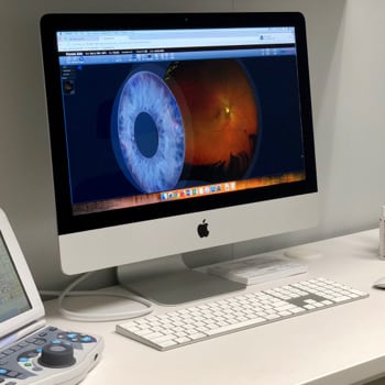
Meibography is a tool that gives your optometrist a much clearer picture of what’s happening behind the scenes. It uses non-invasive infrared imaging to show the structure of the meibomian glands in real time.
What It Helps Detect
This specialized imaging allows your eye doctor to:
- See if the glands are blocked or have begun to atrophy (shrink or disappear)
- Evaluate how many glands are still active and healthy
- Track changes in gland structure over time
- Make better-informed decisions about how to treat your symptoms
With this level of detail, your eye care team can identify meibomian gland dysfunction early, before it leads to more serious or uncomfortable symptoms.
What to Expect During a Meibography
Comfortable, Fast & Informative
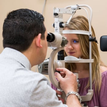
If your optometrist suggests meibography, there’s no need to worry; it’s a quick and comfortable part of your exam.
The Meibography Process Involves:
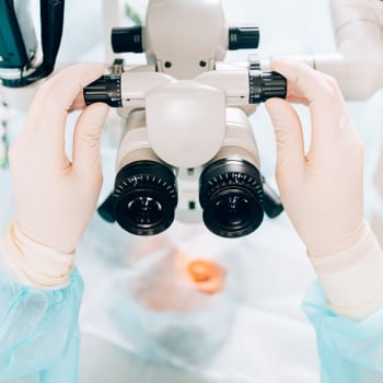
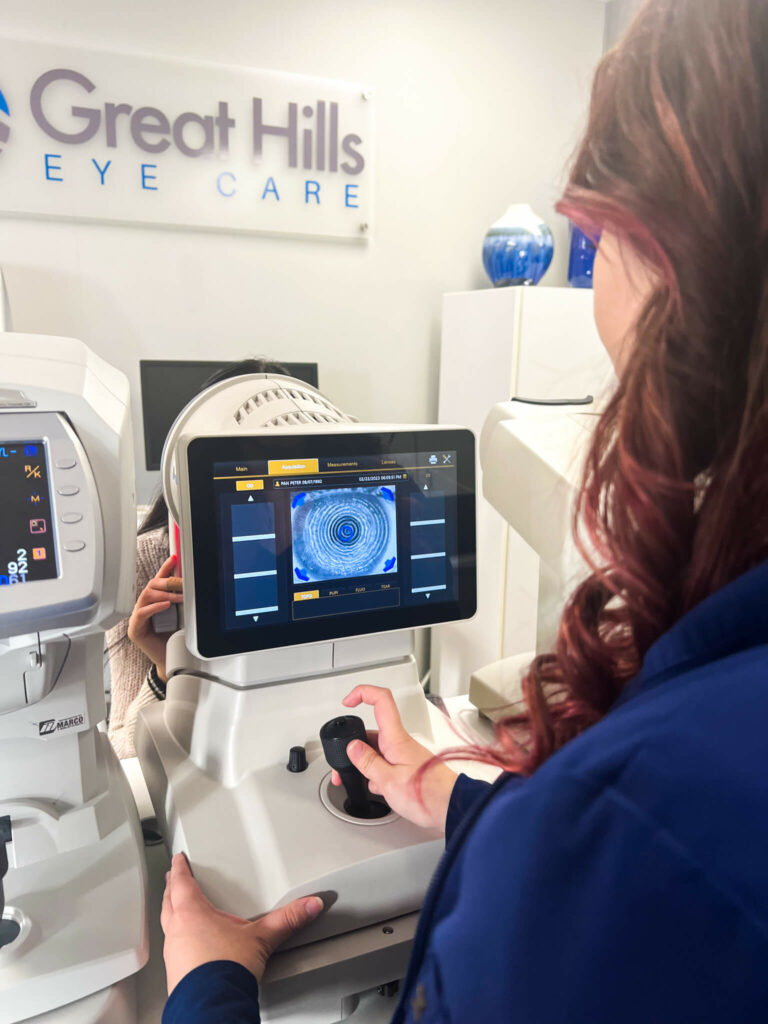
- Sitting in front of a special camera that uses infrared light
- Resting your chin and forehead while the images are taken
- Gently flipping your eyelid to get a clear view of the glands
- No drops, contact, or discomfort during the scan
The entire process takes only a few minutes. Once the images are captured, your optometrist can review them with you, showing exactly how your glands look and explaining what that means for your eye health.
Why Meibography Is Becoming More Common
More Than Just Symptom Relief
As dry eye disease becomes more widely recognized, tools like meibography are becoming essential in eye care practices. The ability to see what’s going on inside the eyelids means that treatment can be more specific and effective.
Rather than relying on symptoms alone, optometrists can build a clearer picture of the root cause. In many cases, this leads to more targeted recommendations; whether that’s lid hygiene practices, warm compresses, prescription drops, or in-office procedures designed to unblock the glands.
Patient Education & Empowerment
It also means patients are more involved in their care. With the ability to see their own gland images, many people gain a better understanding of their condition and feel more empowered to stick to a treatment plan.
Dry Eye & Meibomian Gland Dysfunction
Recognizing the Signs
Not all dry eye is caused by meibomian gland dysfunction, but a large percentage of it is. Understanding the link between the two can help you recognize when to speak up about your symptoms.
Some signs of meibomian gland dysfunction include:
- Burning, stinging, or scratchy eyes
- Sensitivity to light
- Excessive tearing (reflex tears from dryness)
- Blurred vision that improves with blinking
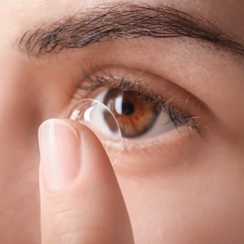
- Discomfort when reading, using screens, or wearing contact lenses
If any of these symptoms sound familiar, meibography might be a helpful part of your exam. It provides clarity not just for your optometrist, but for you as well.
A Better Approach to Dry Eye Care
From General Treatment to Personalized Plans
Meibography fits into a broader shift in how dry eye disease is understood and managed. Where treatment used to rely mostly on symptom relief, newer tools like this support a more proactive and informed approach.
By identifying gland issues early, your optometrist can work with you to help preserve function and comfort. And by tracking changes over time, they can adjust your treatment to fit your evolving needs.
Dry eye management doesn’t have to be one-size-fits-all. With meibography, it can be tailored specifically to the way your eyes work; and what they need to feel more comfortable.
Convenient, Comfortable & Professional Care
Great Hills Eye Care uses meibography as part of a thorough approach to eye exams, especially when dry eye is a concern. The practice is located in the same building as a Costco warehouse but has its own entrance and operates independently, giving patients flexibility to choose where they purchase glasses or contact lenses.
Appointments are available the same day in many cases, and the team prioritizes both comfort and clarity in every visit.
If you’re dealing with dry eyes or have questions about your symptoms, you don’t have to figure it out on your own. Book an eye exam today to find out whether meibography can be part of your personalized care plan.