
Our ability to see is one of the most remarkable ways we engage with the world around us. And safeguarding our vision is just as crucial as maintaining other aspects of our health and well-being.
Optomap is an effective tool for retinal imaging that delivers an accurate view of the eye’s internal anatomy. It plays a vital role in identifying potential problems early and monitoring any changes as they occur over time.
Thanks to technological advancements, we have these types of noninvasive methods to gain insight into parts of our health we can’t typically observe, regardless of how much we might try. Your eye doctor may use a tool like Optomap at your next comprehensive eye examination to provide a detailed perspective on your vision and ocular health.
Understanding Optomap
The retina is a thin layer of light-sensitive tissue on the back of our eyes that plays an essential part in vision and eye health. It detects incoming light and translates it into electrical signals, enabling us to perceive sharp, clear images.
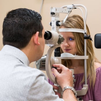
Your eye doctor assesses interior structures like the retina to thoroughly evaluate your eye health. With technology like Optomap, this process has never been more accessible.
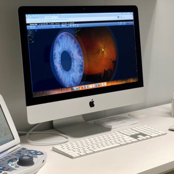
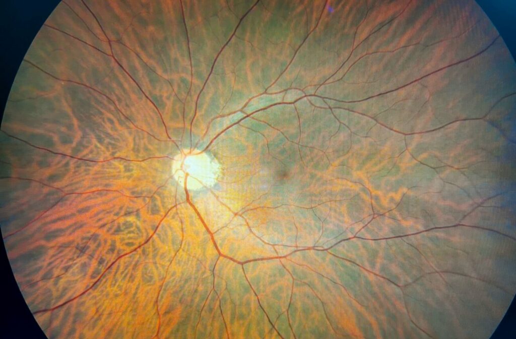
Optomap retinal imaging captures a 200-degree panoramic image of your eye and retina. This expansive view aids your eye doctor in detecting issues in the retina’s peripheral regions.
How Does Optomap Function?
Optomap employs scanning laser technology to take images. It uses two low-power lasers—one red and one green—to scan the back of your eye and produce a high-resolution image of the retina.
These images provide a snapshot of your retinal health and offer a benchmark for monitoring changes over time. The process is fast, simple, and doesn’t require dilation. With just a button press, your optometrist can gain crucial information that might reveal the initial signs of vision-threatening conditions such as glaucoma, macular degeneration, and diabetic retinopathy.
Retinal Imaging & Eye Health
Retinal imaging involves capturing detailed retina images to examine its health and detect abnormalities. This advanced tool assists eye doctors in identifying both eye-specific and general health issues.
Your eye doctor may use an Optomap scanner during various types of eye exams, including:
- Comprehensive eye exams: Your optometrist can establish a baseline measurement of your retina to monitor its health over time.
- Dilated eye exams: If your optometrist suspects more complex issues, dilation may be necessary to widen the pupils and allow a closer inspection of the eye’s internal structures.
- Emergency visits: Retinal images help detect abnormal changes in the eye in cases of sudden symptoms like flashes of light, floaters, or vision loss.
Retinal images act as a diagnostic tool for identifying the earliest stages of eye conditions, such as:
- Glaucoma: Increased eye pressure can lead to peripheral vision loss. Retinal images may show optic nerve damage or cupping, indicating glaucoma.
- Diabetic retinopathy: Complications from diabetes affecting the retina’s blood vessels. Retinal images may reveal leaking blood vessels, retinal swelling, or abnormal vessel growth, suggesting diabetic retinopathy.
- Age-related macular degeneration (AMD): A progressive condition that leads to central vision loss. Indicators include drusen beneath the retina, pigmentary changes in the macula, and progressive thinning or scarring.
With timely action, our team at Great Hills Eye Care can preserve your health and vision, so don’t wait to book an appointment if you think you may be experiencing any of the above issues.
Flashes & Floaters Detected via Retinal Imaging
Flashes manifest as sudden streaks of light in your visual field, often occurring unexpectedly, even in complete darkness. Floaters resemble drifting shadowy shapes across your sight, like squiggly lines or cobwebs.
Floaters become more common as we age due to the vitreous gel becoming more liquid, causing fibers to clump and cast shadows on the retina. This creates the sensation of “floating” in your vision.
Flashes happen when the vitreous tugs on the retina, stimulating light-sensitive cells. This creates an illusion of light that has no external source.
While flashes’ frequency and intensity may vary, they are typically not “normal.” Similarly, while floaters might be harmless, new or significant quantities could point to underlying concerns worth examining.
When to See Your Eye Doctor
Any harm or disturbances to the retina, which processes visual input, compromises sight. New symptoms related to flashes or floaters indicate potentially serious eye conditions like retinal detachments or tears. Signs to watch out for include:
- A sudden rise in floaters or newly visible ones
- Bright, recurring light flashes
- A shadow or “curtain” encroaching on your vision
- Blurred vision or loss of central/peripheral vision
Optomap’s precision and wide-field imaging can help uncover the root cause of these visual disruptions by capturing comprehensive images of abnormal retinal changes.
Invest in Your Vision with Optomap
Your eyes are invaluable, and taking steps to preserve their health is one of the greatest long-term investments you can make. Optomap captures detailed images of your retinal health, setting a strong foundation for proactive vision care.
Connect with our Great Hills Eye Care team to book your next appointment for an eye examination.